SC 2115 Anatomy and Physiology I
Connective and Muscle Tissue
Objectives for Connective and Muscle Tissue:
Ģ Observe under the microscope the major connective tissues found in the body.
Ģ Understand how the structure of connective tissue is related to its function.
Ģ Learn the diversity of this tissue group along with the common characteristics.
Ģ Study the characteristics of loose, dense, elastic, and
reticular connective
tissue, adipose tissue, cartilage, and bone.
Ģ Compare the interrelationship of epithelial and connective
tissue through a
study of the skin.
Ģ Observe the characteristics of the three types of muscle tissue.
Part 1: Overview of Connective Tissue:
This is the most abundant tissue in the body with widespread distribution. The roles of this tissue include support, binding, cushioning, and storage of nutrients. This tissue contains a blood supply, has nerve endings, and repairs body organs. While connective tissues vary widely in structure there are three common characteristics:
A. All connective tissue has an extracellular matrix, cells and fibers are held in place by this ground substance that varies from semi-fluid to solid. The matrix is related to the function of each tissue type. For example, bone has a solid calcium matrix for support.
B. All connective tissues contain protein fibers. The most common fiber type is made of the protein collagen; collagen fibers are flexible and strong. Another fiber type contains the protein elastin; elastic fibers allow stretching or expansion and recoil which is necessary for major blood vessels or the lungs. A third fiber type are reticular fibers which are thin, branched threads that hold loose organs together. Lymph nodes and the spleen need this lattice work support.
C. Another component of connective tissue is sparsely dispersed cells. These vary in name depending upon the tissue.
1. Fibroblasts are star-shaped cells responsible for the production of fibers and the matrix
2. Fibrocytes are mature fibroblasts which maintain connective tissues.
3. Mast cells produce the anticoagulant, heparin, as well as the irritating vasodilator, histamine.
4. Some plasma cells are dispersed in connective tissue and release circulating antibodies.
5. Adipose cells are used as cushions and nutrient storage. Their signet ring shape is due to a large central vacuole filled with triglycerides.
6. Chondrocytes are cells associated with cartilage.
7. Osteocytes are mature bone cells found within the calcium matrix.
Part 2: The Major Connective Tissues
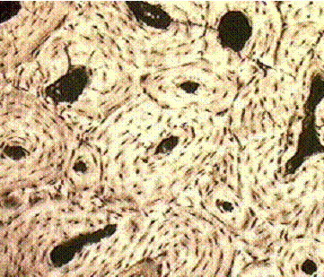
A. Areolar Connective Tissue: packing material for blood vessels and nerves, dermis of skin, and mucous membranes.
This is the most abundant tissue in the body, it covers organs, holds blood vessels and nerves in place, forms the dermis of the skin, and the connective tissue layer of mucous membranes. The matrix is semi-liquid and contains hyaluronic acid which promotes diffusion; it is the dermal layer of the skin which provides nourishment for the epithelial layer. Embedded within the matrix are thick collagen fibers, these are wavy and running in all directions. Thinner, dark elastic fibers form branches and are not as numerous as collagen fibers. The scattered, flat, star-shaped cells are the fibroblasts, while cells containing granules in the cytoplasm are the mast cells.
Draw and label the Areolar Connective Tissue:
collagenous fibers
elastic fibers
fibroblasts
mast cells
B. Dense Connective Tissue: tendons, ligaments, aponeuroses, capsules of organs, and dermis.
There are two divisions: 1) regular dense connective tissue is characterized by collagen fibers held together in parallel bundles with fibroblasts rows sandwiched in between. If you touch your Achilles tendon, you can feel the strong tension that this tissue must withstand. Tendons attach muscle to bone while ligaments hold two bones together at joints. 2) irregular dense connective tissue has thick randomly scattered bundles of collagen fibers with few fibroblasts and little matrix. This tissue serves as the dermis of the skin, the submucosa of the digestive tract, capsules of organs, and tough sheets of fasciae called aponeuroses.
Draw and label White Fibrous Tissue (tendon):
collagenous fiber bundles
fibroblasts
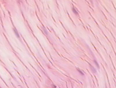
C. Elastic Connective Tissue: walls of arteries and lungs
This tissue contains wavy, thick elastic fibers which appear as though they can stretch as a rubber band. The expansion and recoil of these fibers are associated with air entering and leaving the lungs, with the propulsion of blood through large blood vessels, and with voice production in the vocal cords. When you observe the aorta, locate the parallel wavy branching elastic fibers, a few fibroblasts may be present. In this preparation, there is smooth muscle cells present but not stained.
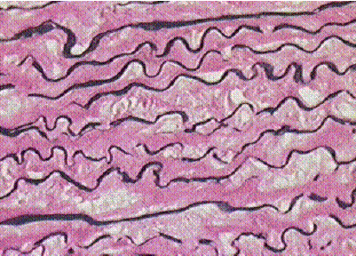
Draw and label the Aorta:
internal elastic fibers
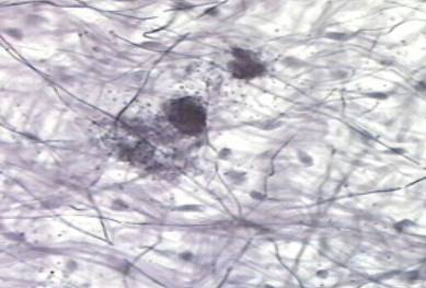
D. Reticular Connective Tissue: forms stroma of liver, spleen, bone marrow, and lymph nodes.
This tissue must be specifically stained and is usually taken from a lymph node or the spleen. These soft organs need an internal scaffolding called the stroma to hold them together. Reticular fibers provide most of the support for the liver and bone marrow as well. The reticular fibers can be described as delicate, heavily-branched, and dark, the matrix is similar to jello.
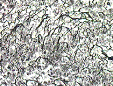
Draw and label Reticular Tissue:
reticular fibers form the stroma
E. Adipose Tissue: surrounds heart and kidneys, subcutaneous tissue, and greater omentum
This is the most easily recognized tissue and will be found widely distributed in every organ microscopically studied this year. The cells are filled with the lipid, triglycerides, and the cytoplasm and nucleus are pushed to the periphery of the cell. Triglycerides are your stored energy, and adipose cells act as good insulators and pillows. There is little matrix associated with adipose tissue but wispy collagenous fibers are threaded between the cells. Fibroblasts present in these fibers can become adipocytes. Adipose tissue is concentrated in the third layer of the skin, the subcutaneous layer, around the heart and kidneys, as yellow bone marrow in long bones, and the bags under oneÆs eyes.
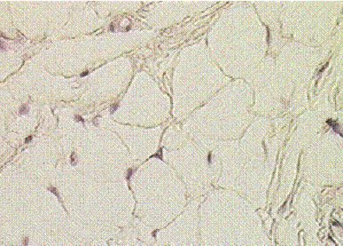
Draw and label Adipose Tissue from the Trachea slide:
adipose cell and nucleus
triglyceride vacuole
collagenous fibers
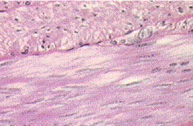
F. Hyaline Cartilage: costal cartilages, articular cartilage,
ends of nose, trachea walls, and
fetal skeleton
This is a tough, glossy, flexible tissue with many functions; it acts as the model for embryonic bone formation and then remains during bone growth as the epiphyseal plate; it is the articular cartilage covering the ends of bones to prevent friction; and it forms the costal cartilages between the first ten ribs and the sternum. Hyaline cartilage prevents the collapse of the trachea as it forms the C-shaped supporting tracheal rings. If you twist the tip of your nose, you can test the resiliency and supportive function of this tissue. Hyaline cartilage has a semi-solid matrix composed of chondroitin which is promoted today as a miracle joint repair. Collagenous fibers are embedded in this matrix but are too thin to be seen. This fiber/matrix composition contains spaces called lacunae which house cartilage cells, chondrocytes. Because cartilage lacks a blood supply, nutrients are received through a membrane surrounding cartilage called the perichondrium. The perichondrium is found in the right side of the picture below at the edge of the oval shaped cartilage.
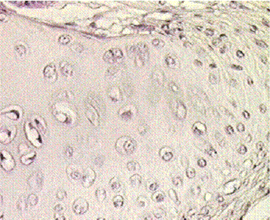
Draw and label the Trachea:
perichondrium (membrane)
fibroblasts in perichondrium
lacuna with 2 chondrocytes
matrix and collagenous fibers
G. Elastic Cartilage: pinna of ear, eustachian tubes, and parts of larynx
If a tissue needs to be pliable yet strong, then adding elastic fibers to hyaline cartilage allows stretching with the return to original shape. The lacunae of elastic cartilage are more prominent and the presence of dark strands of elastic fibers embedded in the matrix.
Draw and label Elastic Cartilage:
lacuna
elastic fibers in matrix
H. Fibrocartilage: pubic symphysis, intervertebral discs, menisci of knee
Many knee problems involve the medial or lateral meniscus, or back problems may involve a slipped disc. The tissue damaged is a fibrocartilage pad. Collagen fibers in fibrocartilage are visible and its matrix and lacunae appear in rows.
I. Compact Bone: periphery of all bones of the skeletal system
Bone is 35% organic matter consisting of cells and collagen fibers and 65% inorganic matrix and calcium salts. There are three cell types associated with bone:
1. osteoblasts: bone forming cells that secrete the matrix
2. osteocytes: mature cells that maintain the calcified matrix
3. osteoclasts: bone destroying cells important in remodeling
Compact bone is the outer layer of dense bone with an organized arrangement called an osteon or Haversian system. Spongy bone is a latticework of porous bone that lies deep to compact bone. Compact bone is hard and dense, its strength comes from osteons or Haversian systems, these are long cylinders arranged parallel to one another along the length of the bone. The Haversian system is arranged in concentric circles of matrix called lamella, in the center is an Haversian canal which houses blood vessels and nerves. In the photomicrograph below, some of the Haversian canals are extended as dark offshoots, these are their interconnections and are called Volkman canals. Note the small nutrient-laden canals called canaliculi radiating from the central Haversian canal, these provide nourishment to osteocytes trapped within the lamellae. The osteocytes are housed within spaces called lacunae.
Draw and label Ground Bone:
haversian system
lamella
lacuna with osteocyte
canaliculi
haversian canal
J. Spongy Bone: underneath the thin layer of compact bone
As the name implies, spongy bone resembles a sponge, that is spicules of bone
called trabeculae which surround marrow cavities. The bone marrow may be red and function in blood cell formation or yellow which contains adipose tissue. In either case spongy bone is highly vascular and well nourished. In the trabeculae, the lamella run parallel and the osteocytes are trapped within lacuna. Spongy bone affords strength with minimal weight. For this drawing you will use developing bone because spongy bone is laid down before compact bone and all three types of bone cells are present.
Within the dark trabeculae are lacuna housing the osteocytes, osteoblasts are found along the developing trabeculae and giant osteoclasts are found near the spongy bone periphery, paticularly in the lower right of this slide. Also visible beyond the periphery is the bone membrane called the periosteum.

Draw and label Developing Bone:
trabeculae with osteocytes
marrow cavity
osteoblasts
osteoclasts
periosteum
Part 3: Observation of the three types of muscle
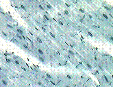
A. Skeletal Muscle: all voluntary muscles attached to the skeleton
This semester you will learn the names of the large skeletal muscles that move bones. A skeletal muscle section under the microscope depicts parallel, elongated cells called fibers with mutliple nuclei at the periphery. Skeletal muscles have a dark and light striations due to the arrangement of the muscle proteins actin and myosin.
Draw and label Skeletal Muscle:
nucleus
striations
skeletal muscle fiber
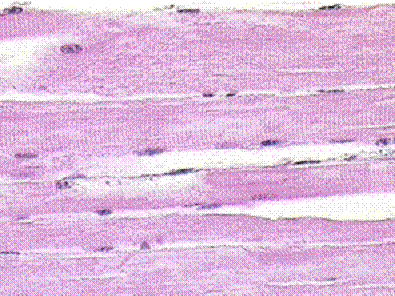
B. Cardiac Muscle: the heart
This muscle type is found only in the heart and is involuntary. The cells have a quadrangular shape with branched ends and a single, central, dark nucleus. Striations due to actin and myosin are present, but the unique feature of these cells is the intercalated disc that is found between two cells. These are distinct, dark bands that allow the cells to communicate quickly so that the cells contract in unison.
Draw and label Intercalated discs, Cardiac Muscle:
striations
nucleus
intercalated discs
C. Smooth Muscle: all involunary muscles associated with viscera
Stomach contrations, peristalsis of the GI tract, blood vessel contriction, goosepumps on the skin, urinary bladder release of urine, and uterine contractions are all involuntarily controlled. Non-striated smooth muscle cells provide the movement of these organs. A smooth muscle cell is a spindle-shaped cell with a central nucleus and tapered ends, these are arranged in bundles so that the tapered ends of one cell fits next to the enlarged center of another cell. In the photomicrograph below, the upper portion is the cross-section of smooth muscle cells and the lower section shows the tapered cells fitting closely together.
Draw and label Stomach Fundus cs: Smooth Muscle:
smooth muscle fibers
nuclei

Test Yourself
1. Describe three characteristics of connective tissue.
a. ___________________________________________________________
b. ___________________________________________________________
c. ___________________________________________________________
2. Match the tissue type in column A with its function in column B:
A B
Connective Tissue Types Function
___areolar a. forms intervertebral discs
___dense b. stroma for soft organs
___elastic c. insulation for the body
___reticular d. shapes the external ear
___adipose e. forms the outer layer of bone
___hyaline cartilage g. provides flexibility to blood vessels
___fibrocartilage h. forms the embryonic skeleton
___elastic cartilage i. attaches muscles to bone as tendons
___compact bone j. soft abundant, packing tissue
___spongy bone k. its spaces are filled with red bone marrow
3. Describe three ways that connective tissue and epithelial tissue are different.
a. ___________________________________________________________
b. ___________________________________________________________
c. ___________________________________________________________
4. Identify the muscle types associated with the following characteristics.
A. voluntary: ____________________________________
B. involuntary: ____________________________________
C. striated: ____________________________________
D. non-striated: ____________________________________
E. multinuceated: ____________________________________
F. single, central nucleus: ____________________________________
G. branched cells: ____________________________________
H. intercalated discs: ____________________________________
I. moves bones: ____________________________________
J. peristalsis: ____________________________________
K. in heart only: ___________________________________