Chapter 23 Notes
Respiratory System
1. Functions
The Air Passageways
Respiratory tract anatomy online
2. Upper Respiratory Tract
1. frontal
2. maxillary
3. sphenoid
4. ethmoid
online sinuses- vestibule contains 3 conchae
- functions to warm air, trap dust, resonation
- surface is pseudostratified columnar epithelium
1. fauces to mouth
2. palatine and lingual tonsils
- laryngopharynx bifurcates into larynx and esophagus
3. Lower Respiratory Tract
1. thyroid cartilage
2. epiglottis
3. cricoid
4. arytenoid- contains vocal cords, these are elastic ligaments that vibrate to produce sound
- muscle stretches ligaments
- air is forced against cords
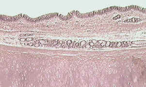
1. mucosa
2. submucosa
3. hyaline cartilage
4. serosa (not shown)
- windpipe with C rings of hyaline cartilage
- anterior to esophagus
- bifurcates into right/left primary bronchi
- bronchial tree online
1. ciliated columnar cells change to cuboidal cells
2. cartilage changes to smooth muscle
Lung Anatomy
4. Lungs
1. base
2. apex
3. costal surface
4. mediastinal surface
5. hilus
6. cardiac notch
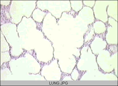
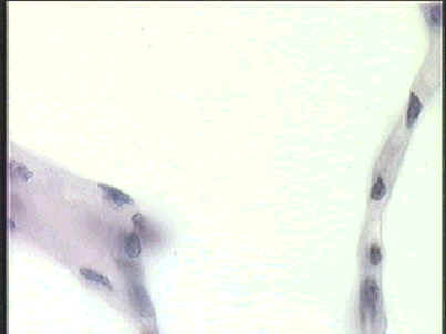
1. Type I cells- for gas exchange
2. Type II cells - secrete surfactant
3. Macrophages - phagocytes
- in emphysema these produce the enzyme
trypsin which destroy the alveolar walls
4. Fibroblasts - secrete elastic fibers
1. gas exchange through a .5 um thick wall
2. note: 2 cells and 2 basement membranes are involved
3. Type I cells, basement membrane, basement membrane, then
capillary cells
4. action: diffusion of gases
Inspiration/Expiration
5. Pulmonary Ventilation
1. Atmospheric Pressure - air pressure
2. Intrapulmonic Pressure - pressure inside the alveoli - varies
3. Intrapleural Pressure -pressure in the pleural space - lower
• INSPIRATION
- Boyle's Law: volume increases, pressure decreases
- diaphragm contracts, dome flattens, phrenic nerve causes this
- ribcage rises and thoracic cavity expands
- intrapulmonic pressure decreases
- air flows from 760 mmHg to 758 mmHg
- muscles of forced inspiration are sternocleidomastoid, pectoralis minor, and
external intercostals
• EXPIRATION
- volume decreases, pressure increases
- diaphragm relaxes, dome shape returns
- rib cage lowers
- intrapulmonic pressure rises to 762 mmHg
- air flows out due to the elastic recoil of the lungs
- lung surfactant reduces the surface tension and causes elastic recoil
- muscles of forced expiration are the abdominals and internal intercostals
6. Compliance - ease of breathing