Chapter 16 Notes
The Special Senses
The
Eye
1. Accessory Structures
- Lacrimal glands:
- - produce tears
- cleanse, lubricate, moisturize
- under parasympathetic nervous system control
- Extrinsic Muscles and cranial nerve control
- - lateral rectus VI
- medial rectus III
- superior oblique IV
- inferior oblique III
- superior rectus III
– inferior rectus III
2. Three Layers of the Eye
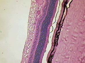
From right to left
1.sclera 2. choroid 3. pigmented epithelium 4. rods and cones
5. nuclear layer 6. layer of synapses 7. bipolar cells 8.
layer of synapses 9. ganglion cells
- Sclera:
- white fibrous connective tissue membrane of eye
- cornea is anterior transparent window V
- Canal of Schlemm is a sinus at junction (drains aqueous humor)
- Choroid
- thin, dark membrane of blood vessels and pigment
- darkens the chamber so light is not reflected
- ciliary body: thickened area of muscle which alters lens shape,
contains ciliary process
which secretes aqueous humor
- iris: colored disk of muscle which controls amount of light, dilation
sympathetic,
constriction parasympathetic
- pupil: black hole which admits light into posterior chamber
- lens: lies behind iris, curved posteriorly held in place by ligaments
from the ciliary body
- Retina
- posterior thin nervous membrane
- extends to ciliary body then ends in a jagged margin called the ora serrata
- contains several layers of neurons- rods, dim light and shape determination
- cones, color vision and bright illumination
- many rods synapse with a bipolar
neurons, only one cone per bipolar
neuron
- ganglion neurons which extend to the Optic Nerve II and exit the eye through
what is called the blind spot
- macula lutea: "yellow spot" 2mm to outer side of optic nerve
and is the exact
center of the retina
- fovea centralis: center of "visual acuity" where images fall,
has high concentration
of cones, no rods
3. Physiology of the Eye
- Refraction
- bending of light as it passes through four transparent media
1. cornea
2. aqueous humor
3. lens
4. vitreous humor
- result is image falls on retina inverted and is
righted by brain
- disorders
1. presbyopia - lens looses elasticity
2. myopia - nearsightedness
3. hypermetropia - farsightedness
4. astigmatism - irregular cornea or lens
- Accommodation |
- lens must change shape to bend light
- ciliary muscle is relaxed, suspensory ligament is taut, the lens is flattened,
light rays are parallel for distant viewing
- ciliary muscle contracts, suspensory ligament slacks, lens bulges for viewing close
objects
- Photopupillary reflex
- iris contracts to constrict pupil and protect retina
- radial muscles dilate the pupil, circular muscles constrict it
- online
colorblind test and another
test
- Visual pathways to the brain
- rods and cones contain photopigments (from Vit. A)
- Rods, rhodopsin is a purple pigment
- Cones: red, blue, green (eat your carrots!)
- Visual pathway online
1. photoreceptors, rods and cones synapse with
2. bipolar cells synapse with
3. ganglion cells
4. impulse travels over optic nerve to
5. optic chiasma, here medial fibers of both eyes cross to
6. optic tract which travels to the thalamus
7. from thalamus through optic radiation to occipital cortex
online
eye
The Ear
in the Temporal Bone
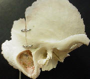
4. External Ear
- Pinna
- elastic cartilage and adipose tissue
- directs sound toward ear
- External Auditory Meatus
- tube in temporal bone
- contains ceruminous glands (secrete wax)
- Tympanic membrane
- fibrous connective tissue
- nerves from mandibular branch of V
5. Middle Ear
- small cavity in petrous portion of temporal bone
- holds 5 drops of fluid
- contains three bones: malleus, incus, and stapes
- vibrations travel over these to oval window
- contains 5 openings
1. to tympanic membrane
2. round window
3. oval window
4. to mastoid sinus
5. eustachian tube
6. Inner Ear
- series of cavities in temporal bone
- vestibule is central portion
- semicircular canals
- project from vestibule posteriorly
- contain fluid called endolymph
- function is equilibrium and balance
- canals contain hair cells which bend as endolymph flows over them,
- VIII is used to send sensory impulses to cerebellum and then cerebral
cortex for skeletal muscle action to maintain equilibrium

- - snail-like organ of hearing
- contains three channels
1. upper scala vestibuli, ends at oval
window contains perilymph
2. middle cochlear duct, contains endolymph
3. lower scali tympani begins at round window contains perilymph
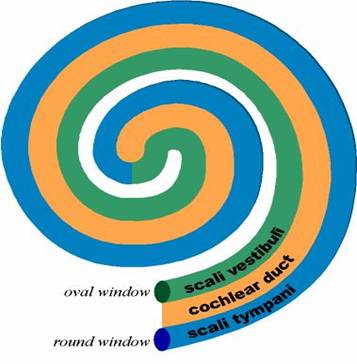
- cochlear
duct is separated from scala vestibuli by the
vestibular membrane,
- cochlear duct is separated from the scala tympani by the
basilar membrane
- resting on the basilar membrane is the organ
of Corti for hearing, this contains hair cells extending into
fluid called endolymph
in cochlear duct, VIII
4. ear antomy online
5. ear
physiology online
7. Physiology of Hearing
- sound waves pass through external ear
- tympanic membrane vibrates
- malleus then incus then stapes vibrate
- stapes pushes on oval window
- a pressure wave forms in perilymph of scala
vestibuli
- wave travels to perilymph in scala tympani
- these pressure waves move the vestibular membrane,
to the endolymph
in the cochlear duct, and finally the basilar membrane
- this bends hair cells on organ of corti bend and
is interpreted by VIII as a sensory impulse
online
ear